Liver Shunt
What You Need to Know About Liver Shunt
When looking for a puppy, one of the greatest concerns that arise is that of Liver Shunt. The following information can be helpful in understanding the types of Liver Shunt and the related conditions and how diagnosis of the disease is detected. Understanding that there is currently no genetic test that can detect whether a dog has the disease or not and what conscientious breeders are doing to help in the eradication of the disease. The following information is compiled from different leading authorities on the disease from the Untied States and the United Kingdom. For more information, visit the Cornell University, University of Florida, University of Tennessee, United Kingdom, and the Yorkie Foundation websites.
Definition and Types
“Portosystemic shunts are abnormal vascular connections between the hepatic portal vein (the blood vessel that connects the gastrointestinal tract with the liver) and the systemic circulation. Such anomalies cause blood in the gastrointestinal track to be diverted past the liver, there by limiting the liver’s vital functions, in metabolism and detoxification of compounds and the body’s defenses against intestinally derived pathogens. This effectively exposes the body to toxic by-products of digestion (toxins and bacteria) and mimics the effects of liver failure.”
T.D.G. Watson, BVM&S, PhD, MRCVS
Waltham Center, United Kingdom
Portosystemic Shunts (PSS) can be classified as Extrahepatic (outside the liver) or Intrahepatic (inside the liver), single or multiple, congenital or acquired.
Extrahepatic Shunts are most common and constitute over 60% of all congenital shunts. Extrahepatic Shunts tend to be found more commonly in small breed dogs, such as Yorkshire Terriers, Maltese, Dachshunds, and Schnauzers. Of these, the Yorkie is 36 times more likely to have congenital Extrahepatic shunts than the other breeds.
Intrahepatic Shunts account for less than 40% of all congenital shunts is more often seen in large breed dogs, such as Irish Wolfhounds and Golden Retrievers. Within the first three days of life the natural opening of the embryonic connection closes. In Intrahepatic Shunts dogs the embryonic connection remains open.
Multiple Acquired Shunts are a result of an increase in liver pressure. This can be a result of the surgical closing of an Extrahepatic Shunt in surgery. Multiple Acquired Shunts are not surgically correctable.
Clinical Sings of Liver Shunt
Dogs with PSS are usually purebred and less than a year old when signs first develop. Poor coat development, lethargy, long sedation recovery, Urinary Tract Infections, disorientation, head pressing, anorexia, depression, weakness, excessive salivating, and temporary blindness have all been connected with Liver Shunt. Vomiting and diarrhea are reported in roughly two-thirds of Liver Shunt cases and in older dogs. Less common signs are recurrent fevers and ascites. Ascites is usually associated only with Acquired Shunts.
Diagnosis of Liver Shunt
In identifying Liver Shunt the identity of the anatomical location of the shunt, its severity, and whether the shunt is congenital or acquired need to be determined. Different Veterinarians utilize different methods for these procedures.
Initially a round of blood work is usually ordered. Common blood work orders are: a full blood chemistry panel, a hematology panel, and a liver profile. It is the combination of elevated liver enzymes combined with low serum cholesterol, hypoglycemia, low blood urea nitrogen, and the total plasma protein concentrations that indicate that a liver shunt is evident.
Serum Bile Acids are also helpful in the making a more definitive overall picture in diagnosing the presence of a Liver Shunt and is usually taken when the blood work is indicative there is a shunt present. It is possible to test the Bile Acids in young puppies. However, unless the puppy is showing apparent signs of PSS a Bile Acid Test can throw false readings. It is recommended an apparently healthy puppy be a minimum of two pounds and 6 to 9 months old to test the Bile Acids accurately.
Once the blood work has been completed multiple testing procedures may take place to help identify the severity and location of the shunt. Three common procedures are: Doppler Ultrasound, Contrast Radiography, and Scintigraphy. Doppler Ultrasound is a 95% accurate and noninvasive procedure for the detection of a Portosystemic Shunts.
Contrast Radiography is the application of a dye injected into the blood stream allowing for easy visualization of the location of the portal shunting and is often done in conjunction with surgery so as to minimize anesthesia of the dog. Scintigraphy is the application of radioactive chemicals into the rectum allowing for the diagnosis to the degree of the shunting allowing a more accurate assessment for management options of the disease.
Treatments and Management of Liver Shunt
Several options exist for dogs diagnosed with Liver Shunt. Many cases can be treated with medical management and diet. Diet is used to limit the production of neurotoxin production in the large intestine thus reducing the stress put on the liver. The limiting of proteins into smaller amounts and only feeding highly digestible sources does this. Protein restricted diets such as Hills, l/d, k/d or Royal Canine Hepatic LS along with lactulose, milk thistle, and occasionally antibiotics are often times successful in treatment of more minor shunt cases.
Surgical options include placements of ameroid constrictors, cellophane bands, and partial suture ligation. 85% of surgical cases of Single Extrahepatic Shunts are successful in the United States. Of the 15% of cases that are not so successful, most are a resulting failure due to either Multiple Acquired Shunts developing from the new pressure being placed on the liver or due a pre-existing condition such as Hypolatic Microvascular Dysplasia or Portal Atresia.
Hypolatic Microvascular Dysplasia (MVD) / Portal Atresia (HMD)
Hypolatic Microvascular Dysplasia or MVD/HMD is a congenital defect where the portal vein that breaks into smaller and smaller microscopic vessels in the liver are poorly formed or undeveloped causing the liver to atrophy which in turn inhibits the liver from filtering out toxins and allowing for normal growth. Dogs can have both a Liver Shunt and MVD/HMD. They can also have one condition without having the other. MVD/HMD livers and shunted liver samples look identical under the microscope and thus can often be misinterpreted for the other. However, a dog with both Liver Shunt and MVD/HMD must have the shunt surgically corrected successfully before a definite conformation of MVD/HMD can be made.
The sign of MVD/HMD are identical to that of Liver Shunt. However, in many cases there are no evident signs of a problem until the dog is 3 to 4 years old. There is currently no treatment for MVD.
Breeding the Future
Hopefully in the future simple blood tests will be able to identify carriers allowing for breeders to eliminate this terrible condition from the Yorkshire Terrier and other dog breeds that are producers of the disease such as Tibetan Spaniels, Cairn Terriers, Havanese, Shih Tzu, and Maltese.
The Yorkshire Terrier Fanciers Foundation is currently working with Dr. Sharon Center, DVM, DiplACVIM, Professor and Internal Medicine Specialist of the department of Clinical Sciences, College of Veterinary Medicine, Cornell University to help provide a base in genotyping Liver Shunt. Dr. Center, as the developer of the Bile Acid Test, believes that Liver Shunt is an ancient mutation in the dog that involves vasculogenesis or angiogenesis (embryologic formation of the blood vessels) and that the need to establish a demonstrated genotype linkage between the breeds is crucial. In the May 2009 Progress Repor to the Canine Health Foundation, Dr. Center reported the determination of Liver Shunt to be an autosonmal dminant but incompletely penetrant mode of transmission and the linkage of the PSVA/MVD trait to a specific chromosome. Progress is being made.
The Foundation has provided a letter of intent to the AKC’s CHF in support of a grant proposal for the project. Dr. Center is currently projecting 18-24 months before a DNA Marker test may become a reality.
As with all genetic and congenital defects, dogs with either Liver Shunt or MVD/HMD should not be used for breeding and should be spayed or neutered.
Genetic Testing and What it Means to the Prospective Buyer
Meanwhile, the dog community anxiously awaits and contributes to the development of a genetic test for MVD and Liver Shunt. As there is currently no genetic test for Liver Shunt, the closest and best test at present is the Serum Bile Acid. All breeding dogs should be tested at Fasting and Post Prandial blood draws. Normal Values are 5 to 15 umol/L with Abnormal Values being over 30 umol/L. Remembering that a dog at a minimum of two pounds and 6 to 9 months old is required to test the Bile Acids accurately, and that the most accurate testing being after a year old when the dog’s liver is fully grown and has had a chance to balance and function properly with the full size of the dog, testing 12 week old puppies before placement into their new homes is not accurate, it can provide a false sense of security to the buyer. However, the Bile Acid test results of both the sire and the dam should be available to the prospective buyer, remembering that the Serum Bile Acids are an overall view of the functioning of the liver, and that is the best that either the breeder or the prospective buyer has to go on until a genetic test is developed and made available to the dog community.
Written by Jennifer White
http://www.vet.utk.edu/clinical/sacs/calendar/
http://www.vetsurgerycentral.com/pss.htm
http://www.yorkiefoundation.org
Luxating Patellas
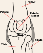
 Luxating Patella (LP) is caused by the rotation of the tibia and the curved
formation of the lower femur resulting in the structural misalignment
of the patella (knee cap) causing slippage out of the trochlear groove
(two bony ridges that form a fairly deep groove in which the patella
is supposed to slide up and down). In a normal dog, the trochlear
groove limits the patella’s movement to one restricted place controlling
the activity of the quadriceps muscle. The entire system is constantly
lubricated by joint fluid allowing for the freedom of motion between
the structures.
LP can be congenital or trauma induced. Females are one and half times more likely to have LP than males, though it is unknown why. LP is progressive and worsens with age as repeat dislocation of the patella causes permanent cartilage damage which can lead to osteoarthritis.
Grades
Luxating Patella is a examination based graded condition rated from 1 to 4. Levels 1 and 2 being relatively minor and manageable to levels 3 and 4 which are more necessitating of surgical correction.
Grade 1: Upon physical examination the patella can be luxated manually. However, the patella does not luxate much on its own generally staying within the trochlear groove.
Grade 2: The patella is easily manipulated during exam out of the trochlear groove and luxations occur when there is occasional spontaneous lameness but the patella returns to normal positioning. This is typically the dog that occasionally carries a rear leg for two or three steps on occasion but then puts it back down and goes as if nothing was wrong.
Grade 3: The patella doesn't always return to normal positioning when it is deliberately pushed out of its groove during a physical examination. Luxation occurs often and the dog has a degree of loss of function due to the luxation. They have more frequent "skipping" episodes, may not want to jump up onto things, and they may have pain.
Grade 4: The leg can not be fully straightened manually and/or the gait is stiff legged due to the patella being underdeveloped or permanently dislocated and fixed in place outside its normal position. The dog shows evidence of chronic pain or disability, including poor to no ability to jump. Luxations are painful enough that the dog tries not to use them. Symptoms include rear lameness, running and screaming in sudden pain as the knee cap dislocates, the holding up of the leg, and the inability to bear weight on the knee.
Surgical Options
There are three types of surgery that can alter both the affected structures and the movement of the patella: block osteotomy or trochlear modification, lateral imbrication, and tibial crest transposition. The type of surgery or combination methods performed depends on the individual case’s cause and severity and the veterinarian.
During block osteotomy or trochlear modification, the trochlear groove may be surgically deepened to better contain the knee cap. This preserves the cartilage block that the patella rides on and creates a equal depth of the groove to prevent further dislocation. During lateral imbrication, the patella itself may be "tied down" laterally (on the outside) to prevent it from deviating medially (toward the inside). If the attachment of the patellar ligament to the tibia, called the tibial crest, is in the wrong position, it is repositioned in a tibial crest transposition. This is done by creating a cut in the tibial crest and reattaching the bone in a position so that the patella is realigned within the trochlear groove. This transposition allows the tendons to be attached in a more lateral position.
There is a 90% success rate with surgical correction. The dog usually is actively using their limb well 2 to 3 months after surgery. Exercise must be restricted for 8 weeks after the surgery or breakdown of the repair may occur. Surgery will not remove the arthritis that may be present in the knee and there maybe some stiffness of the limb or some lameness after heavy exercise.
Due to the complications of surgery on elderly dogs, those with level 3 and 4 LP in their senior years usually have the condition managed with Prednisone. Great strides have been made in the medical management of LP with hydrotherapy which has proven to be a beneficial treatment for low grade LP.
http://en.wikipedia.org/wiki/Luxating_patella
http://en.wikipedia.org/wiki/Canine_hydrotherapy
http://www.veterinarypartner.com/Content.plx?P=A&S=0&C=0&A=2186
http://www.veterinarypartner.com/Content.plx?P=A&S=0&C=0&A=2448
http://www.vetsurgerycentral.com/patella.htm
Poisionous Plants
Acokanthera Aconite (Monkshood) Akee (Soapberry) Aloe Amaryllis Amsinckia (Tarweed) Anenome (Wildflower) Angel Trumpet Tree Anthurium Apple Apricot Pits Arrowhead Vine Asparagus Atamasco Lily Atropa Belladonna Australian Laurel Autumn Crocus Avocado Alligator Pear Azaleas Balsam Pea Baneberry (Doll's Eyes) Beach Pea Betel Nut Palm Belladonna Bird of Paradise Bittersweet Black Locust Black Walnut Bleeding Heart Bluebonnets Blue Flag (Iris) Blue-Green algae Bloodroot Bottlebrush Bouncing Bet (Soapwort) Boxwood Braken Fern Buckeye Horse Chestnut Blackthorn Broad Bean Buttercup Caladium Calendula (Marigold and other names) Calla Lilly Canary Bird Bush Candelabra Cactus Cardinal Flower Carolina Jessamine Cashew Cassava Castor Bean Castor-Oil Plant Cat Claw Catelaw Acacia Ceriman Chalice Vine (Trumpet Vine) Cherry (Wild and Cultivated) Cherry Laurel Chinaberry Tree Chinese Evergreen Choke Cherry Christmas Berry Christmas Cactus Christmas Candle Christmas Rose Chrysanthemum Climbing Lily Coffee Weed Columbine Common Burdock Common Cocklebur Common Privet Coral Plant Corn Cockle Corn Lily Coyotillo Crinum Lily Crocus Croton Cotoneaster Cowslip Cursed Crowfoot Cyclamen Daffodil Daphne Datura (Jimson Weed) Deadly Amanita Deadly Nightshade Death Camas Death Cap Mushroom Delphinium Dieffenbachia (Dumb Cane) Destroying Angel (Death Cap) Dogwood Dumb Cane (Dieffenbachia) Dutchman's Breeches Easter Lily Eggplant Elderberry Elephant Ears English Holly English Ivy Equisetum |
Eucalyptus Euonymus Euphorbic (Crown of Thorns, Spurge) European Beech Evening Trumpet Flower False Hellebore False Henbane Fava Bean Fiddleneck (Senecio) Fig (Ficus) Flax Fly Agaric (Amanita, Death Cap) Four O’clock Foxglove Gelsemium Ghost Weed Glory Lily Golden Chain Golden Dewdrop Green Dragon Heliebore Heliotrope Hemlock (Poison & Water) Henbane Holly (English & American) Horse Bean Horse Chestnut Horsetail Reed (Equisetum) Hyacinth Hydrangea Impatiens Indian Hemp (Dogbane) Indian Tobacco Indian Turnip(Jack-in-the-Pulpit) Inkberry Ink weed (Dry Mary) Iris(Blue Flag) Ivy (All Forms) Jack-in-the-Pulpit Japanese Yew Jasmine (Yellow) Jasmine (Star) Jatropha Java Bean Jerusalem Cherry Jessamine Jimson Weed (Thorn Apple) Johnson Grass Jonquil Juniper Kentucky Coffee Tree Klamath Weed Laburnum Lady's Slipper Lambkill (Sheep Laurel) Lantana Camara Larkspur Laurels Lily of the Valley Lima Bean (Java Bean) Lobelia Locoweed Locust Lord and Ladies (Cuckoopint) Lupine Machineel Magnolia Maidenhair Tree (Ginko Biloba) Mandrake Marijuana Marsh Marigold May Apple Mescal (Bean) Mexican Bird of Paradise Mexican Breadfruit Mexican Tea Milkweed Mistletoe Moccasin flower (Lady Slipper) Mock Orange Mole Bean Monkshood Moonseed Morning Glory Mountain Laurel Mushrooms and Toadstools (Wild Types) Mustard Narcissus (Paper-White, Daffodil) Natal Cherry Nectarine Seed Nepthytis Nettle Nicotiana (Wild and Cultivated) Night-Blooming Jessamine Nightshades Narcissus (Paper-White, Daffodil) Natal Cherry Nectarine Seed Nepthytis Nettle Nicotiana (Wild and Cultivated) Night-Blooming Jessamine |
Nightshades Pasque Flower Pathos Paw Paw Peach Pear Pennyroyal Peony Periwinkle Peyote Pheasant's Eye Philodendron Pigeonberry Pigweed Pinks (Sweet William, Carnation) Pittosporum Plums Poinsettia Poison Hemlock Poison Ivy Poison Oak Poison Sumac Pokeweed/Poke cherry Poppy (except California) Potato Pothos Precatory Bean Primrose PRIMULA Privet Ragwort Ranunculus Rattlebox Red Oak Rhododendron Rhubarb Ricnus Rosary Peas Rose Bay Rosemary Rubbervine Russian Thistle Sage Salmonberry Scarlet Pimpernel Scotch Bloom Senecia (Fiddle Neck) Silver Leaf Night Shade Skunk Cabbage Sky Flower Duranta Snapdragon Sneezeweed Snowdrop Sorghum Sour Dock (Sorrel) Spanish Bayonet Spider Lily Spindle Tree Spurges Star Jasmine Sudan Grass Star of Bethlehem (Snowdrop, Nap-at-Noon) Stranomium St. John's Wort Sundew Swamp Lily Sweet Pea Tansy Taro (Elephant Ears) Tarweed Texas Mountain Laurel Thorn Apple Tiger Lily Toad Flax Tobacco Tomato Plant Touch-Me-Not Toyon Berry Trillium Tri-leaf Wonder Trumpet Vine Tulip Venus Fly Trap Verbena Vetches Virginia Creeper Walnut Water Hemlock White Snakeroot (Richweed) Wild Black Cherry (Choke Cherry, Rum Cherry) Wild Cucumber (Manroot) Wild Onion (also cultivated onion) Wild Parsnip Wisteria Wormseed Yam Bean Yellow Oleander Yellow Star Thistle Yew (American and English) Yucca Plant Zephyranthes Lily |
Additional Links
www.yorkiehealthfoundation.org
www.cfytc.org/health/issues.htm
